Test Complete
- Questions
- Score
- Minutes
| Overall Results | |
|---|---|
| Total Questions |
| Category Results | |
|---|---|
Cardiovascular Anatomy Basics
Category: Cardiology
Topic: Cardiovascular Anatomy and Physiology
Level: EMT
Next Unit: Blood Vessels
18 minute read
Basic Cardiovascular Terminology
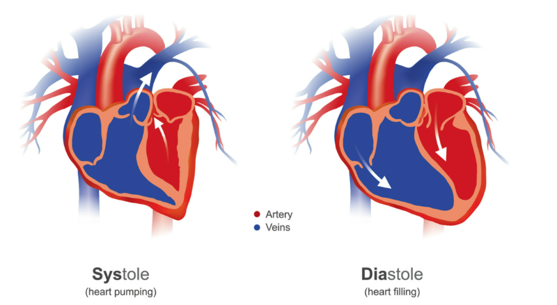
CARDIAC CYCLE: The cardiac cycle is made up of two phases, systole and diastole.
- Systole is when the heartbeat contracts and pumps blood from the chambers into the arteries.
- Diastole is when the heart muscle relaxes and allows the chambers to fill with blood.
PULSES: The body presents with two types of pulses, peripheral and central.
- Peripheral pulses are found in the extremities.
- Central pulses are located directly off the carotid or subclavian arteries (i.e. the neck).
BLOOD PRESSURE: The blood pressure is quantified by two numbers in a fraction representation (e.g., 120/80):
TOP NUMBER (Systolic): refers to the amount of pressure in your arteries during contraction of your heart muscle.
BOTTOM NUMBER (Diastolic): refers to the amount of pressure in your arteries during the relaxed period between beats. The diastolic pressure is sometimes referred to as the end-diastolic pressure, indicative of the baseline vascular resistance in the body's tissues.
SYSTOLE: The "heartbeat" happens in two sequential contractions, one of which sends blood from the atria into the ventricles and another, slightly skewed in its timing, that sends the received blood out of the ventricles to the body. An EKG demonstrates this well, with the P-wave representative of atrial contraction and the QRS representative of ventricular contraction.
CARDIAC OUTPUT: the product of heart rate and blood volume ejected per beat (stroke volume).
CO = HR x SV.
PERFUSION: The measure of the saturation of tissues with oxygenated blood.
It is dependent upon correctly functioning red blood cells that carry oxygen-saturated hemoglobin to the tissues and cells of the body, as well as sufficient rate, pump (contraction force), and volume.
OXYGENATION of Tissues: Homeostatic blood flow delivers oxygenated blood and removes tissue wastes.
General Anatomy and Function
The heart is, in essence, a double organ:
- It is a heart that takes deoxygenated blood and pumps it to the lungs for oxygenation.
- It is also a heart that pumps the oxygenated blood throughout the rest of the body, including the arteries that supply itself.
There are 4 chambers:
RIGHT SIDE of the HEART
The RIGHT ATRIUM pumps deoxygenated blood to the RIGHT VENTRICLE.
From the right ventricle, the blood is pumped to the lungs via the pulmonary arteries. Returning from the lungs where oxygenation took place, the oxygen-rich blood is drawn into the LEFT ATRIUM.
LEFT SIDE of the HEART
The LEFT ATRIUM receives oxygenated blood from the lungs and pumps it into the LEFT VENTRICLE through the mitral valve.
The LEFT VENTRICLE then pumps this oxygen-rich blood into the aorta, the main artery that carries blood to the entire body, including the head and neck regions.
HEART VALVES: Backflow Prevention
The heart is a pump. Contractions of cardiac muscle constrict (contract) the chambers and the pressure within them increases, causing the blood to flow forward through paths of least resistance.
The flow from each atrium to its corresponding ventricle is a one-way flow, with backflow prevented by valves that open in only one direction, slamming shut to any pressure that would try to send blood backward ("retrograde"). These atrioventricular valves divide each atrium from its corresponding ventricle. Likewise, the outflow from each ventricle is guarded against backflow by the semilunar valves.
The heart has papillary muscles that connect to the cusps of the atrioventricular valves (tricuspid and bicuspid valves), via chordae tendinae and prevent the inversion or prolapse of these valves upon systole.
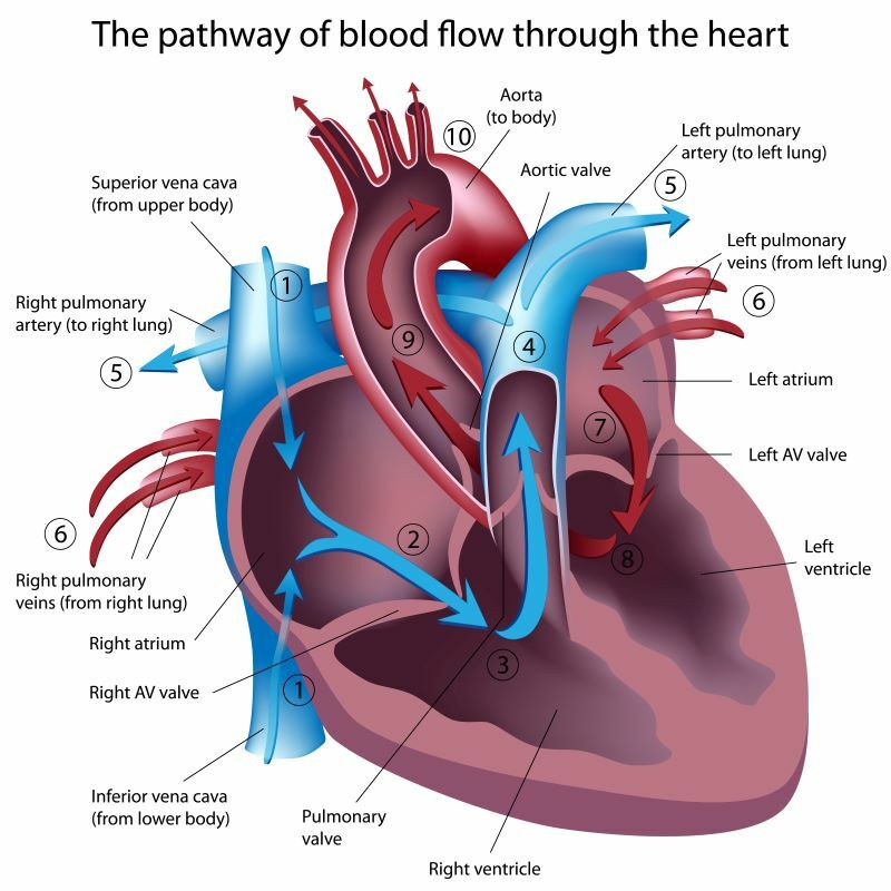
ITINERARY:
Right Atrium → [through tricuspid atrioventricular valve] → Right Ventricle → [through Pulmonary semilunar valve]
[⇒LUNGS⇒]
Left Atrium → [through mitral "bicuspid" atrioventricular valve] → Left Ventricle → [through Aortic (bicuspid, or semilunar) valve] → REST OF THE BODY + THE HEART ITSELF (Coronary Arteries)
Therefore,
- the atrioventricular valves (R-tricuspid and L-bicuspid or mitral) keep blood from flowing back into the atria, and
- the R-pulmonary semilunar and L-aortic bicuspid semilunar valves prevent blood from flowing back into the ventricles.
- The pulmonary valve separates the right ventricle from the pulmonary circulation.
- The aortic valve prevents blood from pumping from the aortic arch back into the left ventricle.
To illustrate: when the heart contracts, the increased pressure within
the right ventricle forces the blood backward toward a closed tricuspid valve and forward through an open pulmonic valve toward the lungs. The closed tricuspid valve prevents backflow, and the one-way pulmonic valve blows open by the passing of blood.
Likewise, the left ventricle forces the blood backward toward a closed mitral valve and forward through an open one-way aortic valve toward the aortic arch.
INFLOW
The blood that comes to the right atrium comes from the
- Superior Vena Cava above, and the
- Inferior Vena Cava below.
By the time these large veins dump into the right atrium, they have already had contributions from the liver veins (the Portal System) and the lymphatic ducts.
The blood that returns from the lungs gets to the left atrium via the Pulmonary Veins.
Don't get confused: this arrangement, besides fetal circulation, is the only one in which arteries are oxygen-poor (pulmonary arteries) and the veins are oxygen-rich (pulmonic veins).
OUTFLOW
- The upper body, neck, and head are supplied by the Aortic Arch. After the aortic arch branches off into arteries supplying these parts of the body, it continues on as
- the Descending Thoracic Aorta, after passing through the diaphragm, continues farther as the Abdominal Aorta to supply the chest, abdomen, and lower body.
- The heart itself is supplied by two major coronary arteries that come off the beginning of the aortic arch right beyond the aortic valve.
Generally, the left side ventricular pressure is higher than the right side ventricular pressure.
The Heart as a Pacemaker
The heart is an unusual muscle in that it has a structure called the Sino-Atrial Node (SA, or sinus node). It regularly fires off nerve conduction bursts on its own but is also under modification by inputs from the autonomous nervous system through both the sympathetic ("fight-or-flight") and parasympathetic ("rest-and-digest") nervous systems.
The sympathetic fibers innervate the ventricles to increase heart rate and force of contraction; the parasympathetic (vagus origin) target the SA node to inhibit heart rate and force of contraction.
Normal Sinus Rhythm: generally, the 60-100 beats/minute via the SA node. The SA node is in the right atrium, near where the superior vena cava enters.
To act as an effective pump, it is important that all 4 chambers aren't contracting at exactly the same time. Therefore, the spread of electrical stimulation originating in the SA node allows a sequential contraction sequence among the 4 chambers that generally moves
RIGHT → LEFT and SUPERIOR → INFERIOR
From the SA node, branches travel this R → L and inf → sup route.
From the SA node, there are nerve branches to the L atrium and down to the atrioventricular (AV) node, and from there down the Bundle of HIS, from which branches then go on to spread throughout the ventricles as the Purkinje Fibers.
SUCCINCTLY: SA node → AV node → HIS Bundle → Purkinje Fibers. All of this takes time:
Admittedly, not a lot, but enough to stagger the contractions such that the vector flow of blood is in the OUT OF THE HEART direction!