Test Complete
- Questions
- Score
- Minutes
| Overall Results | |
|---|---|
| Total Questions |
| Category Results | |
|---|---|
Blood Vessels
Category: Cardiology
Topic: Cardiovascular Anatomy and Physiology
Level: AEMT
Next Unit: The Venous System
20 minute read
Blood vessels function to transport blood. Generally, they distribute oxygen-rich blood to the tissues via arteries and their branches and collect de-oxygenated blood via the venous (vein) system of veins, called tributaries, not branches.
Confluence, like in bodies of water, is the meeting together of tributaries. Divergence, like from the trunk of a tree, is a branching out. Therefore, arteries create branches and veins are created from tributaries.
Hydrostatic Pressure within Blood Vessels
If you put your thumb over the spout of a garden hose, occluding it, the continued water flow will build up the pressure within the hose. If that same amount of water were in a hose that had a larger diameter, the pressure would be lower than the pressure of that water in a thinner hose. Likewise, the diameter of a vessel influences the pressure of the blood within it.
Hydrostatic Pressure: the pressure that blood exerts against the walls of the vessel.
Oncotic Pressure: the force that tends to pull water into the circulatory system and prevents it from leaving.
These two forces are opposing and interact to result in the distribution of blood throughout the body.
Round Trip
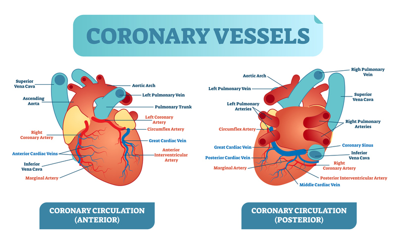
When oxygenated blood leaves the left ventricle, it travels through the ascending aorta and the descending (thoracic and abdominal) aorta, into the branching arteries, then into smaller arterioles, and finally into capillaries.
In the capillaries, gaseous exchange with cells takes place, and then deoxygenated blood travels from the capillaries to the venules, then to the veins, then to the superior and inferior vena cava and back into the heart by way of the right atrium.
From the right atrium, the blood is sent to the right ventricle, and from the right ventricle, (still deoxygenated) blood travels through the pulmonary arteries to the lungs, where it is oxygenated, and then through the pulmonary veins to the left atrium, and finally to the left ventricle again to repeat the cycle.
Collateral Circulation
The blood supply to many parts of the body have redundancies built in that allow for continued oxygenation even if supplying branches of arteries become blocked.
An example of this is the uterus, which has blood flow coming from the uterine arteries, but also has blood supply (collateral circulation) coming from above it, from branches of the ovarian arteries.
The Circle of Willis is a circular arterial anastomosis (the connection or joining of two normally separate tubular structures) located at the base of the brain. It connects the internal carotid arteries with the vertebrobasilar system through a network that includes the anterior cerebral arteries, anterior communicating artery, posterior cerebral arteries, and posterior communicating arteries. This network provides collateral circulation so that blood flow to the brain can continue even if one major artery is narrowed or blocked.
Knowledge of collateral circulation is crucial in surgery to prevent blood loss when only the primary blood supply has been interrupted before removing an organ. Also, collateral circulation explains why applying pressure to the radial artery may not stop the hand from bleeding, due to collateral circulation from the ulnar artery and the middle palmar arterial arches.
Not all organs or tissues enjoy the advantages of collateral circulation, and these are particularly at risk to cell death/organ failure if a sole blood supply is impaired in some way. For example, blood supply to the colon is at risk from an embolism that blocks the middle mesenteric artery.
IN A NUTSHELL:
L ventricle → (ascending/descending aorta) → arteries → arterioles → capillaries →
[GASEOUS EXCHANGE: Cellular Respiration (glucose + oxygen → water + carbon dioxide )]
→ venules → veins → inferior/superior vena cava → R atrium → R ventricle → pulmonary arteries → LUNGS →
[OXYGENATION/REMOVAL OF CO2 (passive diffusion)]
→ pulmonary veins → L atrium → L ventricle.
Blood Vessel Anatomy
Blood vessels are made up of different layers:
- INTIMA
The Tunica Intima is the innermost layer of the arteries and veins, lines the entire circulatory system and is composed of a thin layer of endothelial cells. Capillaries are only made up of tunica intima, which allows the exchange of gasses and nutrients to take place.
Plaque build-up from elevated LDL-lipoprotein involvement of the intima occurs in atherosclerosis.
- MEDIA
The Tunica Media is the muscular middle layer of arteries and veins. The tunica media of arteries contain more smooth muscle than that of veins, allowing for the arteries to constrict and dilate based on need and on influences from the autonomic nervous system; with releases of epinephrine, vasoconstriction occurs, and with releases of histamine, vasodilation occurs.
- ADVENTITIA
The Tunica Adventitia is the outermost layer of a blood vessel and is mainly composed of collagen, and in arteries, receives extra support from elastic lamina. This collagen acts as an anchor to steady the blood vessel against nearby organs. In general, arteries carry more blood and at a higher pressure than veins do, and capillaries are the least pressurized and have the lowest blood volume.
Between arterial and venous anatomy, there are differences in these three layers. The most dramatic difference is between the Tunica Media of arteries and veins: this layer is thicker in arteries than in veins.
Types of vessel wall injury:
- Longitudinal: tears, lacerations, or crushing along the long axis of the vessel.
- Stretched: resulting in irregular tears when the elasticity is overwhelmed.
- Transverse: when a vessel is cut completely, leaving the injury a circle in cross-section.
By the architectural intactness of the circumferential tunica media in vessels (the encircling muscular layer that contracts or relaxes for vasoconstriction or vasodilation, respectively) that has been completely transected, retraction and vasoconstriction of the vessel will limit or even stop hemorrhage. A longitudinal, stretching, or crushing injury does not leave the circumferential nature of this circular function intact.
- Crushing: all wall integrity of the vessel is destroyed.
Capillary Gas Exchange
Most blood vessels have 3 layers (tunics):
- the Intima (endothelium),
- the Media, and
- the Adventitia.
Capillaries are the interface between the smallest branches of the arterial system 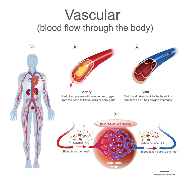 (arterioles) and the smallest tributaries of the venous system (venules), and that position makes them the site of gaseous exchange between CO2 from the veins and O2 from the arteries at the tissue cells where they meet. (Such an exchange is similar--but opposite--in the alveoli of the lungs)
Capillaries are much smaller and thinner than arteries and veins. The walls of these vessels consist of only a single layer of endothelial cells, the Tunica Intima, allowing substances to pass through them quickly.
SIMPLE DIFFUSION: During homeostasis, gasses exchange between blood and cells at the capillary level through the process of simple diffusion. Oxygen-rich blood travels from the arteries to the capillaries surrounding a specific tissue. At the end of the connection, the oxygen-rich blood reaches a point where only a single one-cell-thick wall separates the capillary and the tissue.
Concentration gradient difference between the oxygen-rich blood and oxygen-depleted tissue cells allows for the oxygen to diffuse or pass through the single-cell-thick wall, and into the tissue, in an attempt to equalize the gradient on both sides. The same thing happens for a waste product like carbon dioxide. The carbon dioxide-rich tissue cells, when in contact with carbon dioxide-depleted capillaries, diffuse the area of higher concentration to the area of lower concentration, in an attempt to equalize, causing the carbon dioxide to diffuse through the single-cell-thick wall and into the blood to be excreted from the body elsewhere.
Anastomosis
A circulatory anastomosis is a connection between two blood vessels, such as those between two arteries (arterio-arterial anastomosis), those between veins (veno-venous anastomosis) or those between an artery and a vein (arterio-venous anastomosis). An arteriovenous anastomosis is artificially constructed in the arm for purposes of dialysis.