Test Complete
- Questions
- Score
- Minutes
| Overall Results | |
|---|---|
| Total Questions |
| Category Results | |
|---|---|
Basic Blood Chemistry
Basic Blood Chemistry
Laboratory evaluation of blood provides valuable information about the health of sick or injured patients. By analyzing the concentrations of electrolytes, gases, and other chemicals in the blood, healthcare providers can gain insights into the underlying causes of vague symptoms such as weakness, syncope, or dizziness. While not always available in the prehospital setting, lab values provide EMS with context to anticipate complications and identify high-risk conditions, supporting timely recognition and clearer communication with receiving facilities.
Analysis of cardiac enzymes in the patient's blood can confirm a suspected myocardial infarction or secure the suspicion of strokes.
Cardiac Biomarkers ("Cardiac Enzymes")
Purpose:
Cardiac enzymes are chemicals released by the heart when myocardial cells are injured. Elevated levels of certain cardiac enzymes indicate myocardial damage. Traditionally, the evaluation of cardiac enzymes has been limited to hospital settings. However, with the development of rapid enzyme testing kits, it is now possible to assess some cardiac enzymes in prehospital settings quickly.
Indications:
Many interventions performed in the field can alter blood composition and obscure critical diagnostic information. Having the capability to draw blood before administering treatments allows emergency departments to evaluate a patient's original status and monitor changes over time.
Procedure:
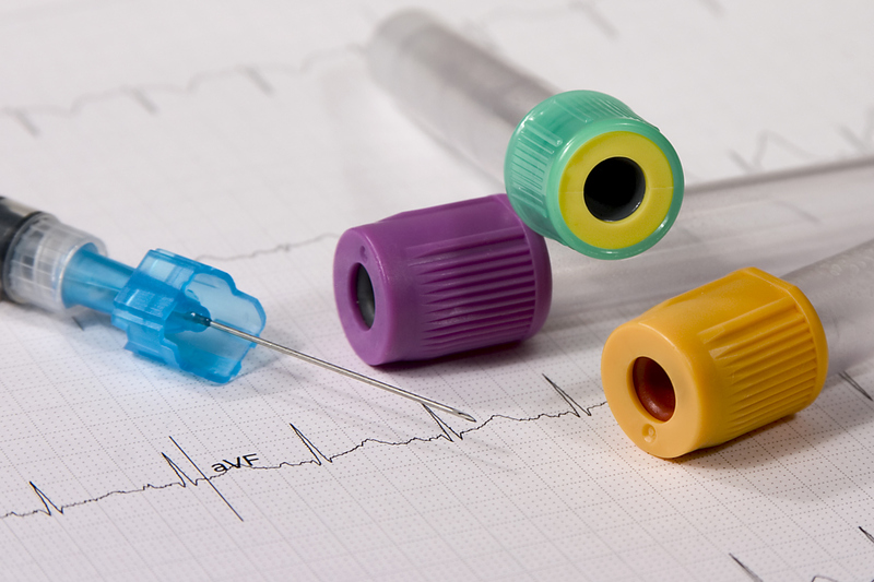
Venous blood is typically obtained via venipuncture. Responders can draw blood samples simultaneously when establishing IV access. Blood samples should be obtained in the following situations:
- During peripheral access (IV)
- Before administering medication
- When medication administration may be anticipated
Important Note: Never delay urgent patient care to perform a blood draw.
Procedures for Blood Draws
- Obtaining Blood from an Angiocatheter:
- Prepare all necessary equipment.
- Check tubes for expiration date and any damage.
- Do not attach tubes to a vacutainer until ready for use to avoid losing the vacuum.
- Establish IV access with the angiocath, but do not connect the IV tubing yet.
- Attach the end of the multi-draw needle adapter to the angiocath hub.
- Insert blood tubes in the correct order, allowing the rubber-covered needle to puncture the tube's self-sealing top.
- Fill all tubes completely, then gently agitate them to mix the anticoagulant with the blood.
- Remove the vacutainer and multi-draw needle, and then attach the IV tubing to ensure patency.
- Label all tubes with necessary patient information.
- Using a 20 mL Syringe:
- Prepare all required equipment.
- Check tubes for expiration date and any damage.
- Attach the syringe needle adapter to the angiocatheter hub and gently withdraw blood.
- Once the syringe is full, remove it from the angiocath and place the IV line.
- Attach a hypodermic needle to the syringe and puncture the tops of the blood tubes.
- Insert blood tubes in the correct order, allowing the needle to puncture the self-sealing top.
- Fill tubes completely and gently agitate to mix the anticoagulant.
- Dispose of sharps safely.
- Label all tubes with the necessary information.
- Direct Venous Blood Draw:
- Prepare all necessary equipment.
- Check tubes for expiration date and damage.
- Do not attach tubes to a vacutainer until ready for use.
- Apply a constricting band and select the venipuncture site, then cleanse the site.
- Insert the Luer sampling needle into the vein, then remove the constricting band.
- Insert blood tubes in the correct order, allowing the needle to puncture the self-sealing top.
- Fill tubes completely, agitate gently, and then apply sterile gauze over the site.
- Remove the needle, dispose of it properly, and cover the puncture site.
- Label all tubes with necessary patient information.
COMPLICATIONS:
Blood draws can lead to complications such as:
- Damage to the vein wall: Caused by incorrect needle placement or movement.
- Hemoconcentration: Occurs when the constricting band is left on too long, leading to an artificially high concentration of red and white blood cells in the sample.
- Hemolysis: The destruction of red blood cells, which can release hemoglobin and potassium into the sample, making it unusable. Hemolysis can be caused by shaking the tubes too vigorously, using a needle that is too small, or forcefully aspirating blood into or out of the syringe.
Common Blood Analyses
- CHEM-7 Panel:
This test measures the levels of seven components in the blood, often used to assess a patient's overall metabolic and renal function:- Sodium (Na+): A key electrolyte that helps regulate fluid balance, nerve function, and muscle contraction.
- Normal range: 135-145 mEq/L
- Abnormal values: Hyponatremia (low sodium) may indicate fluid overload, heart failure, or kidney disease. Hypernatremia (high sodium) may suggest dehydration or endocrine disorders.
- Potassium (K+): Essential for muscle function, including heart muscle, and nerve transmission.
- Normal range: 3.5-5.0 mEq/L
- Abnormal values: Hypokalemia (low potassium) can cause muscle weakness and arrhythmias. Hyperkalemia (high potassium) is dangerous and can lead to cardiac arrest.
- Chloride (Cl-): Works with sodium to maintain fluid and acid-base balance.
- Normal range: 98-106 mEq/L
- Abnormal values: Hypochloremia may occur with vomiting or metabolic alkalosis. Hyperchloremia can be associated with dehydration and metabolic acidosis.
- Bicarbonate (HCO3-) or CO2: Helps maintain the acid-base balance in the blood.
- Normal range: 22-28 mEq/L
- Abnormal values: Low bicarbonate may indicate metabolic acidosis, while high levels may suggest metabolic alkalosis or respiratory compensation for chronic lung disease.
- Blood Urea Nitrogen (BUN): A measure of kidney function and hydration status, reflecting protein metabolism.
- Normal range: 7-20 mg/dL
- Abnormal values: Elevated BUN may indicate kidney dysfunction, dehydration, or high protein intake. Low BUN can be seen in liver disease or malnutrition.
- Creatinine: A byproduct of muscle metabolism, creatinine levels are a more specific marker of kidney function.
- Normal range: 0.6-1.2 mg/dL
- Abnormal values: Elevated creatinine suggests impaired kidney function or acute kidney injury.
- Glucose: A primary energy source for the body, blood glucose levels reflect metabolic and endocrine function.
- Normal range (fasting): 70-100 mg/dL
- Abnormal values: Hypoglycemia (low glucose) can cause weakness, confusion, or seizures. Hyperglycemia (high glucose) is indicative of diabetes mellitus or stress response.
- Sodium (Na+): A key electrolyte that helps regulate fluid balance, nerve function, and muscle contraction.
- Creatine Kinase (CK or CPK):
CK is an enzyme found in the heart, brain, and skeletal muscle. Elevated CK levels typically indicate muscle damage. CK-MB, a specific form of CK, is more specific for myocardial injury.- Normal range: 22-198 U/L (varies based on gender and age)
- Abnormal values: Elevated CK-MB levels can suggest a myocardial infarction, with levels rising 4-6 hours after the event and peaking at 12-24 hours.
- Lactic Dehydrogenase (LD or LDH):
LDH is an enzyme found in various body tissues, including the heart, liver, kidneys, and muscles. It is released into the bloodstream during tissue damage.- Normal range: 140-280 U/L (varies based on laboratory standards)
- Abnormal values: Elevated LDH levels may indicate myocardial infarction, hemolytic anemia, liver disease, or certain cancers. It begins to rise within 24 hours of an MI and returns to normal within 2 weeks.
- Myoglobin:
Myoglobin is a protein found in cardiac and skeletal muscles that binds oxygen. It is one of the first markers to rise after muscle injury.- Normal range: 0-85 ng/mL
- Abnormal values: Myoglobin is one of the first markers to rise after muscle injury. It begins to rise within 1-2 hours after a myocardial infarction (MI), peaks at 6-8 hours, and returns to normal within 20-36 hours. Myoglobin is less specific than troponin because it can also be elevated in skeletal muscle injuries.
- Troponin:
Troponin is a regulatory protein in heart muscle fibers. It is the preferred biomarker for detecting myocardial infarction due to its high specificity for cardiac tissue damage.- Normal range: Troponin I <0.04 ng/mL, Troponin T <0.01 ng/mL
- Abnormal values: Troponin is the preferred biomarker for detecting myocardial infarction due to its high specificity for cardiac tissue damage. Troponin may begin to rise within 2-4 hours post-MI. Troponin levels peak at 12-16 hours and can remain elevated for up to 10 days, making it more reliable for diagnosing and assessing the extent of myocardial injury over time.
- B-Natriuretic Peptide (BNP):
BNP is a hormone produced by the heart's ventricles in response to increased pressure and volume overload, commonly seen in heart failure. It helps to differentiate between cardiac and non-cardiac causes of dyspnea.- Normal range: Less than 100 pg/mL
- Abnormal values: Elevated BNP levels (greater than 100 pg/mL) suggest heart failure. Levels above 400 pg/mL are strongly indicative of congestive heart failure (CHF). The higher the BNP level, the more severe the heart failure.
- Arterial Blood Gas (ABG):
ABG analysis provides detailed information about a patient’s oxygenation, ventilation, and acid-base status. It is critical in evaluating patients with respiratory distress, metabolic disorders, or those requiring intensive care. ABGs are typically drawn from the radial artery but can also be obtained from other arterial sites.- Components measured:
- pH: Measures the acidity or alkalinity of the blood.
- Normal range: 7.35-7.45
- Abnormal values: pH below 7.35 indicates acidosis, while pH above 7.45 indicates alkalosis.
- Partial Pressure of Oxygen (PaO2): Reflects the amount of oxygen gas in the blood.
- Normal range: 75-100 mmHg
- Abnormal values: PaO2 less than 60 mmHg indicates hypoxemia.
- Partial Pressure of Carbon Dioxide (PaCO2): Indicates the effectiveness of ventilation.
- Normal range: 35-45 mmHg
- Abnormal values: PaCO2 above 45 mmHg suggests respiratory acidosis (hypoventilation), while below 35 mmHg indicates respiratory alkalosis (hyperventilation).
- Bicarbonate (HCO3-): Acts as a buffer to maintain normal pH levels.
- Normal range: 22-28 mEq/L
- Abnormal values: Low bicarbonate may indicate metabolic acidosis, and high bicarbonate may indicate metabolic alkalosis.
- Oxygen Saturation (SaO2): Percentage of hemoglobin saturated with oxygen.
- Normal range: 94-100%
- Abnormal values: SaO2 less than 90% often indicates the need for supplemental oxygen.
- pH: Measures the acidity or alkalinity of the blood.
- Components measured: