Test Complete
- Questions
- Score
- Minutes
| Overall Results | |
|---|---|
| Total Questions |
| Category Results | |
|---|---|
Basic Airway Anatomy
Category: Airway
Topic: Airway Anatomy
Level: EMR
Next Unit: Structures of the Airway
18 minute read
Basic Airway Anatomy
Upper Airway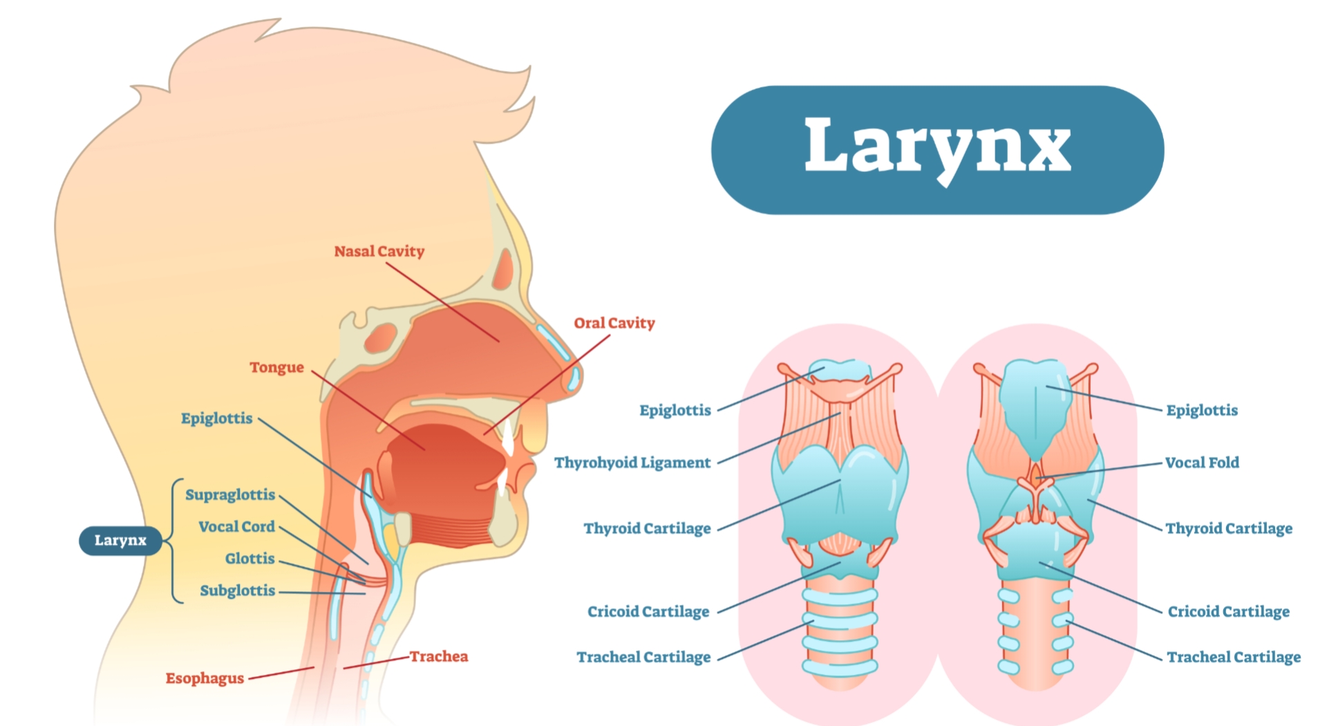
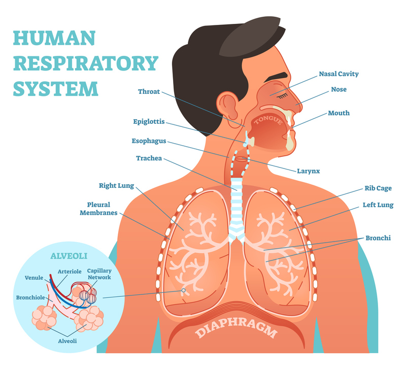
The upper airway is the "A" of the ABCs. As the entry point for oxygen any damage to, or blockage of, the structures in the upper airway can rapidly result in unconsciousness or death. The anatomy of the upper airway can be broken down into the nose, mouth, and throat. The medical terms for these are the nasopharynx and oropharynx/larynx.
NOSE (Nasopharynx): The nose is the primary airway used by most conscious adults to breathe. The space behind the nostrils (the nasopharynx) is filled with blood-rich tissue covered in mucus, which warms and cleans incoming air.
MOUTH: The mouth is used as an alternate airway in normal adults and is especially important in emergency situations, when the nasal pathway may be blocked due to illness or trauma. The mouth is also the entrance to the digestive system and is involved in the production of speech.
THROAT (Oropharynx/Larynx): The oropharynx is the area behind the tongue at the very back of the mouth, it connects to the nasopharynx superiorly and the larynx inferiorly. The larynx and oropharynx are separated by the epiglottis.
- The Larynx is where sound is produced. Vibrations of cartilage and tissue caused by fast-moving air lead to sound, which is formed into speech by the tongue and mouth.
- The Epiglottis is the mechanism that covers the opening of the trachea when food is swallowed. It acts as a "trap door" which closes when swallowing to prevent food from entering the lower airway.
Lower Airway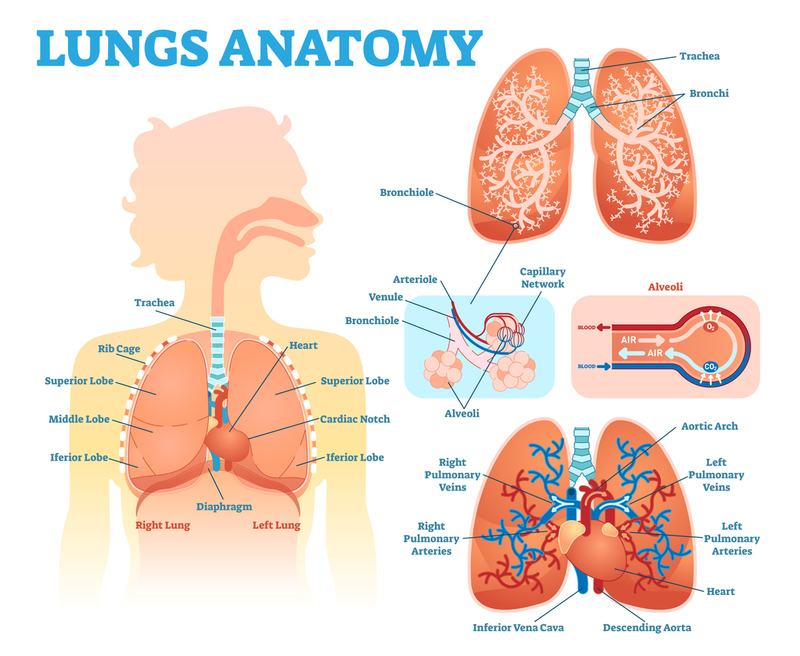
The lower airway is made up of all structures below the larynx (voice box). As in the upper airway, there are structures that transfer air; the trachea, bronchi, and bronchioles, and structures that allow oxygen and carbon dioxide to be exchanged with the blood, the alveoli.
TRACHEA: The trachea is a hollow tube that passes air to the lower airways, it differs from the airway structures above it in that it is supported by cartilage rings. The trachea sits anterior to the esophagus.
BRONCHI: The bronchi are hollow tubes that branch off of the trachea at the carina into right and left bronchi, these are known as the main stem bronchi. They then further subdivide into smaller bronchi for each lobe of the right and left lung. These structures are supported by cartilage rings.
The right mainstem bronchus points downward at a sharper angle than the left. This is why foreign bodies or aspirated material are more likely to get stuck in, or pass through, the right mainstem bronchus.
BRONCHIOLES: The bronchioles are smaller than even the bronchi and lie between the bronchi and the alveoli, they differ from the bronchi in that they do not have cartilage rings, and stay open via smooth muscle.
ALVEOLI: The alveoli are millions of microscopic, thin-walled sacs located deep within the lungs. These sacs are surrounded by a network of capillaries, which allow for efficient exchange of gases. Oxygen is taken in by the alveoli and passed through the capillaries to the rest of the body, while carbon dioxide, a waste product of cellular metabolism, is picked up by the capillaries and exhaled out of the body. The alveoli are the terminal point of the respiratory system and play a vital role in maintaining the body's oxygen levels and removing carbon dioxide.
Breathing (Ventilation vs. Respiration)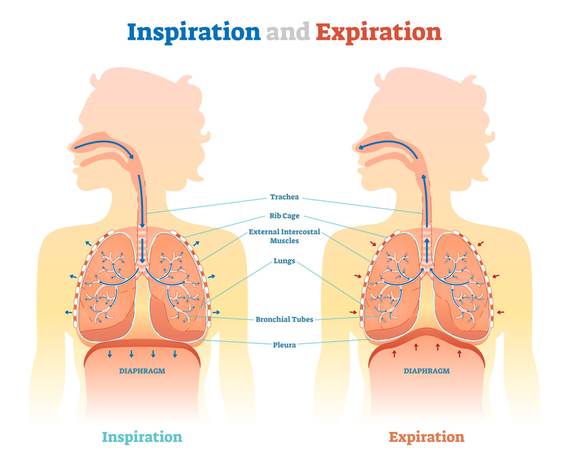
DEFINITIONS
- Ventilation: The inspiration and expiration of air due to lungs filling and emptying via the movement of the diaphragm and intercostal muscles.
- Respiration: The exchange of oxygen and carbon dioxide. External respiration occurs in between the outside air and the blood. Internal respiration occurs between the blood and the cells.
- Carina: The point where the trachea splits into the left and right mainstem bronchi. The approximate location of the carina on the outside of the body can be seen at the Angle of Louis (manubriosternal joint) on the chest, approximately at the level of the second pair of ribs.
In summary, ventilation is the movement of air in and out of the lungs, while respiration is the exchange of gases between the blood and another environment. Keep these definitions in mind as you may be asked to differentiate between the two on exams.
The pathway of air during ventilation is as follows:
nose → nasopharynx → glottis → trachea → main stem bronchi → bronchioles → alveoli.
The pathway of blood flow (for respiration) is as follows:
right heart -> pulmonary arteries -> alveolar capillaries -> pulmonary veins -> left heart -> body
It is important to remember that the pulmonary arteries carry deoxygenated blood and the pulmonary veins carry oxygenated blood, this is the opposite of the arteries and veins in the rest of the body and is commonly tested.
Musculoskeletal Cage
THORACIC CAGE: The thoracic cage is made up of ribs (12 on each side) joined to the sternum anteriorly and wrapping around posteriorly to the spine. The rigidity provided by the ribs is vital to ventilation, as the diaphragm would not be able to draw air into the lungs without a solid structure to pull against. 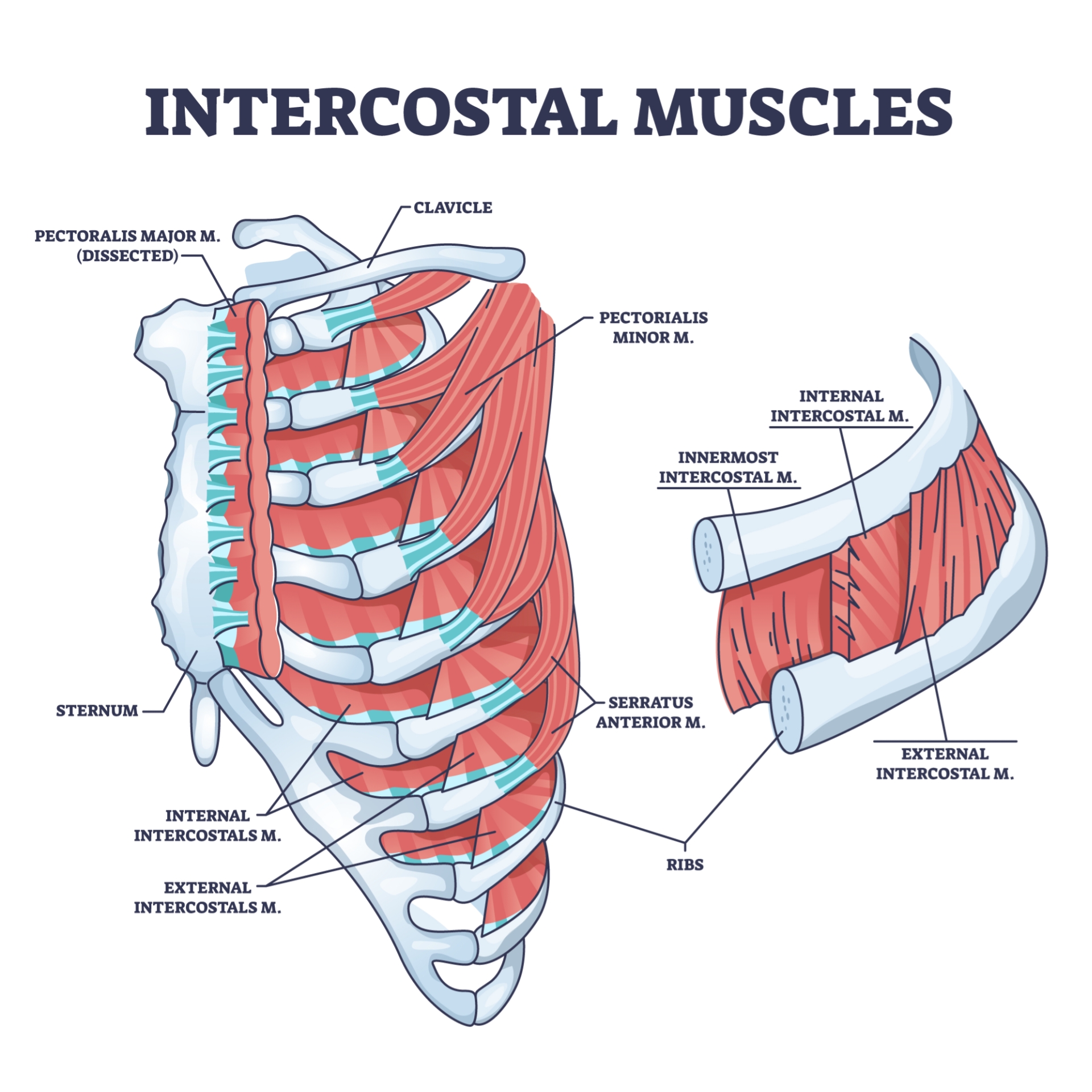
MUSCLES OF RESPIRATION: The muscles of respiration include the muscles between the ribs (intercostals) and the diaphragm, which is the primary muscle of respiration.
The intercostal muscles lie between the ribs and pull the ribs together, helping to shrink the size of the chest cavity and making the exhalation of air easier. Along with the ribs, they form the chest wall, which protects the organs.
The neck and back muscles also assist with respiration, but they help with inspiration in cases where massive airflow is needed, not during normal respiration at rest.
The diaphragm is the main ventilatory muscle. When it contracts, it pulls itself downward, creating negative pressure in the chest, which causes air to rush into the lungs. When it relaxes, it is pulled upwards by the lungs natural recoil. This causes air to rush out of the lungs like a collapsing balloon.
The diaphragm is controlled by both the autonomic and voluntary nervous systems. This allows the respiratory rate to vary automatically based on the blood concentrations of CO2 and Oxygen that the brainstem senses while also allowing you to speed up or slow your respiratory rate at will. All nerve signals to the diaphragm go through the phrenic nerve, which comes from the nerve roots of the cervical spine.
The Mediastinum
The mediastinum is the center of the thoracic cavity, which lies between the two lungs containing several structures. Those involved in the respiratory system are the; heart, trachea, and great vessels (Aorta, pulmonary vessels, vena cava).